基于CT序列图像的肺部管道三维分割方法的研究
VIP免费
I
摘要
在所有癌症中,肺癌的发病率和死亡率都居于前列,是危及人类生命健康的
“头号杀手”。肺癌的早期诊断和治疗是提高肺癌患者存活率的关键。由于早期肺
癌一般以肺结节的形态存在,因此,对肺结节的检测就成了肺癌早期诊断的主要
手段。
目前,对肺结节检测最为有效的手段是多层螺旋 CT。由于多层螺旋 CT 对肺
部扫描的图像数据巨大,人工检测肺结节不仅效率低,而且容易造成漏诊、误诊。
计算机辅助检测(computer-aided diagnosis, CAD)技术可以大大提高肺结节的诊断
准确率与工作效率,减少漏诊误诊。CAD 是目前生物医学工程领域的研究的热点
之一。
基于 CT 图像,实现对肺部管道(气管树及血管树)的3D 分割是肺结节计算机
辅助检测技术研究中不可缺少的步骤。在 CAD 系统中,对分类器进行训练所使用
的特征主要是灰度特征、纹理特征和几何特征(包括 2D 和3D 特征)。2D 几何特
征主要是:圆形度、面积、线形度及边界的形状等特征。在 2D 情况下,肺结节与
血管和气管在形态上相似,但在 3D 情况下,由于气管和血管的特征表现为管状结
构(Tube-like),结节的特征通常表现为球形结构(Spherical)。因此,能否提取出
肺部血管和气管的 3D 特征,对区分结节与血管等不同组织至关重要,这就要求从
肺部 CT 图像中准确提取出血管和气管的 3D 结构。通过分析血管/气管和结节的
3D 形态特征,一方面可以在一定程度上降低结节检测的假阳性率,另一方面可以
进一步确定结节的位置,提高结节检测的准确度。
分割肺气管主要有三个方面的困难。首先,随着气管逐渐分级、变细,其管
壁变薄,气管内腔 CT 值和管壁 CT 值差异变小;其次,由于部分容积效应的影
响,气管的边界变得比较模糊。最后,当气管走向与 CT 断层扫描的截面方向平
行时,其管腔不能完全被管壁包围。由于上述困难的存在,在分割时容易产生气
管断裂及误分割现象。
分割肺血管的主要问题:首先,在肺部 CT 图像中,肺血管对比度低、结构错
综复杂;其次,图像的部分容积效应、病人呼吸等原因造成的成像伪影; 最后,
高密度气管壁容易与血管混淆。以上问题给肺部血管的分割造成了一定困难。
针对上述问题,本文以准确分割肺部管道组织为目的,提出了一种基于阈值、
三维区域生长、形态学分割及线状结构组织滤波器的综合分割方法。实验结果证
明,本文提出的方法能够快速、自动、准确地分割肺气管;对于肺血管分割的初
II
步检测,本文的方法能够取得较好的分割效果。今后,经过进一步的算法学习和
改进,相信会取得更好的分割效果。从而为后续肺结节计算机辅助检测技术的研
究奠定了良好的基础。
本文主要研究工作体现在以下几个方面:
(1) 提出基于阈值及三维连通域标记的肺实质分割方法。该方法直接对三维数
据进行处理,减少了循环处理单幅二维图像的繁琐,能够简单有效的提取肺实质
区域。另外,该方法提出应用三维区域生长方法去除大气管,并实现了种子点的
自动选取。
(2) 分别利用三维区域生长及形态学方法进行肺气管分割。通过分析分割结
果,对比其优缺点,提出一种改进的区域生长方法---结合形态学与区域生长的分
割方法。首先,利用区域生长法初步提取气管树,其次,利用形态学分割方法选
取候选细小气管区域;最后,利用区域生长法提取出最终的气管树。
(3) 研究了基于 Hessian Matrix 的管道组织滤波器的设计,通过对血管增强图
像进行阈值操作初步获取肺血管。
关键词:CT 序列图像 管道分割 三维连通域标记 三维区域生长 形态
学分割 Hessian 矩阵
III
ABSTRACT
In recent years, lung cancer is the primary cause of cancer related deaths. In the
early period, lung cancer generally exists in the form of pulmonary nodules. Therefore,
early detection of potentially cancerous pulmonary nodules may be a key way to im-
prove a patient’s chances for survival.
Computed tomography (CT) is the most commonly used imaging modality for de-
tecting small pulmonary nodules because of its sensitivity and ability to repre-
sent/visualize a completely three-dimensional (3D) structure of the human thorax.
However, each scan contains numerous sectional images that must be evaluated by a
radiologist in a potentially fatiguing process. As a result, the performance of a human
interpreter can be adversely affected. It is therefore an important task to develop com-
puter aided detection (CAD) systems that can aid/enhance radiologist workflow and po-
tentially reduce false negative findings.
Pulmonary vascular tree and airway tree segmentation is the indispensable step of
the computer aided detection of pulmonary nodules. In CAD systems, the main features
used in training the classifier are gray features, texture features and geometric features,
including 2D and 3D features. The main features of 2D geometry are circular, area, lin-
earity, and boundary shape. In 2D images, it’s similar between lung nodules and blood
vessels. But in 3D images, the vascular and airway are tube-like, while the nodules are
spherical-like. Therefore,it’s essential to extract the 3D features of the pipe structures
(vascular tree and airway tree)for distinguishing different structures such as nodules and
vessels. There are two functions to extract the 3D features of the pipe structures. On one
hand, it potentially reduces false negative rate of nodule detection. On the other hand, it
can further determine the location of nodules which improves the nodule detection ac-
curacy.
There are three difficulties in airway segmentation. First, with the trachea gradual-
ly grading, thinning, the difference of CT value between the tracheal wall and cavity
become smaller. Second, the boundaries become more blurred because of the partial
volume effects. Finally, the trachea lumen cannot be completely surrounded by its walls
when it parallels the image. Because of these difficulties, it’s prone to the lung bronchial
tube rupture and leakage.
IV
There are also three problems in pulmonary vascular segmentation. First, in CT
images, pulmonary vascular usually have low contrast and structural complexity. Se-
cond, it always produces artifacts caused by partial volume effects and patient’s breath-
ing. Finally, high density airway walls can be confused with vessels.
Our presented methods are based on threshold, three-dimensional region growing,
morphological segmentation and vessel filter. Experimental results show that our meth-
ods are rapid, automatic and accurate for the airway segmentation. As the initial seg-
mentation of pulmonary vessels, the experimental results show that the method pro-
posed in this paper could obtain good segmentation results. Also, we could make better
segmentation effect after further study and improvement of the algorithms in future.
Therefore, it makes a good foundation to the follow-up study of pulmonary nodules
CAD.
The key points of the research are as follows:
1. The paper presents 3D lung segmentation method based on threshold and 3D
components labeling. It produces 3D data directly, reducing the complexity of
processing 2D images one by one. In addition, it proposed a 3D region growing
method which can select seeds automatically.
2. In this paper, a combined method was proposed. First, using 3D region growing
to segment the main airway. Second, using the morphology method to segment
the small regions. Finally, extract the final tree by using region growing again.
3. Study a vessel filter based on hessian matrix—Hessian filter and Frangi’s filter.
And use thresholding to segment the pulmonary vascular.
Key Word :CT Image Sequence, Pipeline Segmentation,
Three-Dimensional Connected Labeling, Three-Dimensional Region
Growing, Morphological Segmentation
V
目录
摘要
ABSTRACT
第一章 绪论 .................................................................................................................... 1
§1.1 引言 ................................................................................................................ 1
§1.2 本文主要研究内容 ........................................................................................ 3
§1.3 本文结构 ........................................................................................................ 3
第二章 医学图像分割方法概述 .................................................................................... 5
§2.1 图像分割概述 ................................................................................................ 5
§2.2 图像分割定义 ................................................................................................ 6
§2.3 图像分割方法分类 ........................................................................................ 6
§2.4 三维(3D)图像分割概述 ................................................................................ 9
§2.5 图像分割方法评价 ...................................................................................... 10
§2.6 小结 .............................................................................................................. 10
第三章 基于肺部 CT 序列图像的肺实质的三维分割 ............................................... 12
§3.1 引言 .............................................................................................................. 12
§3.2 医学图像处理平台 ...................................................................................... 13
§3.3 肺部 CT 图像预处理 ................................................................................... 15
§3.3.1 加权均值平滑滤波 ......................................................................... 15
§3.3.2 自适应中值滤波 ............................................................................. 16
§3.3.3 形态学基础 ..................................................................................... 17
§3.3.4 形态学滤波 ..................................................................................... 18
§3.3.5 实验结果与分析 ............................................................................. 18
§3.4 三维肺实质自动分割 .................................................................................. 19
§3.4.1 二值化处理 ..................................................................................... 20
§3.4.2 三维连通域标记 ............................................................................. 21
§3.4.3 大气管的提取 ................................................................................. 22
§3.4.4 肺实质的获取 ................................................................................. 23
§3.5 实验结果与分析 .......................................................................................... 23
§3.6 小结 .............................................................................................................. 25
VI
第四章 肺气管的分割 ................................................................................................ 26
§4.1 引言 .............................................................................................................. 26
§4.2 区域生长算法原理 ...................................................................................... 28
§4.2.1 区域生长算法 ................................................................................. 28
§4.2.2 区域生长算法类型 ......................................................................... 29
§4.2.3 区域生长准则 ................................................................................. 30
§4.2.4 像素的邻域 ..................................................................................... 31
§4.2.5 像素的连通性 ................................................................................. 32
§4.3 区域生长算法程序设计 .............................................................................. 33
§4.4 基于形态学的分割 ...................................................................................... 34
§4.4.1 二值形态学 ..................................................................................... 35
§4.4.2 灰度形态学 ..................................................................................... 36
§4.4.3 基于形态学的气管树提取 ............................................................. 38
§4.5 实验结果与分析 .......................................................................................... 39
§4.5.1 实验数据获取及实验环境 ............................................................. 39
§4.5.2 基于区域生长的分割 ..................................................................... 39
§4.5.3 基于形态学的分割 ......................................................................... 39
§4.5.4 基于形态学及区域生长的分割 ..................................................... 41
§4.5.5 结果与讨论 ..................................................................................... 42
§4.7 小结 .............................................................................................................. 44
第五章 肺血管分割的初步研究 ................................................................................ 45
§5.1 引言 .............................................................................................................. 45
§5.2 Hessian 矩阵定义 ......................................................................................... 46
§5.3 Hessian 矩阵应用 ......................................................................................... 47
§5.3.1 Hessian 矩阵与向量函数的极值 ..................................................... 47
§5.3.2 Hessian 矩阵用于三维图像的增强滤波器 ..................................... 48
§5.3.3 Hessian 矩阵用于肺血管检测 ......................................................... 49
§5.4 肺血管提取 .................................................................................................. 50
§5.5 实验结果与分析 .......................................................................................... 50
§5.5.1 实验数据获取 ................................................................................. 50
§5.5.2 实验结果分析 ................................................................................. 51
§5.6 小结 .............................................................................................................. 54
第六章 结论与展望 .................................................................................................... 55
VII
§6.1 结论 .............................................................................................................. 56
§6.2 展望 .............................................................................................................. 56
参考文献 ........................................................................................................................ 58
在读期间公开发表的论文和参加的科研项目 ............................................................ 63
致 谢 ............................................................................................................................ 64
相关推荐
-
七年级数学下册(易错30题专练)(沪教版)-第13章 相交线 平行线(原卷版)VIP免费
 2024-10-14 42
 -
七年级数学下册(易错30题专练)(沪教版)-第13章 相交线 平行线(解析版)VIP免费
 2024-10-14 56
 -
七年级数学下册(易错30题专练)(沪教版)-第12章 实数(原卷版)VIP免费
 2024-10-14 40
 -
七年级数学下册(易错30题专练)(沪教版)-第12章 实数(解析版)VIP免费
 2024-10-14 31
 -
七年级数学下册(压轴30题专练)(沪教版)-第15章平面直角坐标系(原卷版)VIP免费
 2024-10-14 53
 -
七年级数学下册(压轴30题专练)(沪教版)-第15章平面直角坐标系(解析版)VIP免费

 2024-10-14 45
 -
七年级数学下册(压轴30题专练)(沪教版)-第14章三角形(原卷版)VIP免费

 2024-10-14 41
 -
七年级数学下册(压轴30题专练)(沪教版)-第14章三角形(解析版)VIP免费
 2024-10-14 49
 -
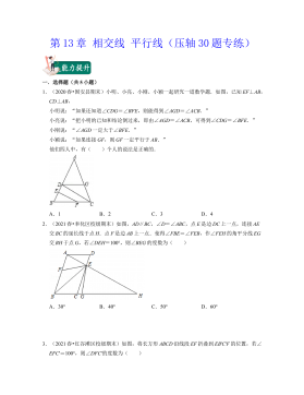
七年级数学下册(压轴30题专练)(沪教版)-第13章 相交线 平行线(原卷版)VIP免费
 2024-10-14 45
 -
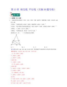
七年级数学下册(压轴30题专练)(沪教版)-第13章 相交线 平行线(解析版)VIP免费
 2024-10-14 45
作者:陈辉
分类:高等教育资料
价格:15积分
属性:69 页
大小:3.39MB
格式:PDF
时间:2025-01-09
相关内容
-

七年级数学下册(压轴30题专练)(沪教版)-第15章平面直角坐标系(解析版)
分类:中小学教育资料
时间:2024-10-14
标签:无
格式:DOCX
价格:15 积分
-

七年级数学下册(压轴30题专练)(沪教版)-第14章三角形(原卷版)
分类:中小学教育资料
时间:2024-10-14
标签:无
格式:DOCX
价格:15 积分
-

七年级数学下册(压轴30题专练)(沪教版)-第14章三角形(解析版)
分类:中小学教育资料
时间:2024-10-14
标签:无
格式:DOCX
价格:15 积分
-

七年级数学下册(压轴30题专练)(沪教版)-第13章 相交线 平行线(原卷版)
分类:中小学教育资料
时间:2024-10-14
标签:无
格式:DOCX
价格:15 积分
-

七年级数学下册(压轴30题专练)(沪教版)-第13章 相交线 平行线(解析版)
分类:中小学教育资料
时间:2024-10-14
标签:无
格式:DOCX
价格:15 积分


